

 Posted By Akio
- Comments 0

What Is the Retina of the Eye?
Your retina is a thin layer of tissue at the back of your eye.
A simple way to understand it is:
- The cornea and lens focus light.
- The retina detects and processes that light.
- The optic nerve carries visual information to the brain.
- The brain interprets the signals as the images you see.
What does the retina do?
- Detects light entering your eye
- Converts light into electrical signals
- Sends those signals to your brain through the optic nerve
- Processes colour, motion, and fine detail
How Do Rods and Cones Help You See?
The retina contains two main types of photoreceptor cells: rods and cones.
Rods
Rods are highly sensitive to low levels of light. They support:
- Night vision
- Peripheral or side vision
- Detection of movement
- Vision in dim environments
They are found predominantly outside the centre of the retina.
Cones
Cones function best in brighter light and support:
- Colour vision
- Fine detail
- Reading
- Face recognition
- Sharp central vision
Cones are highly concentrated in the fovea, the central part of the macula responsible for the clearest vision.
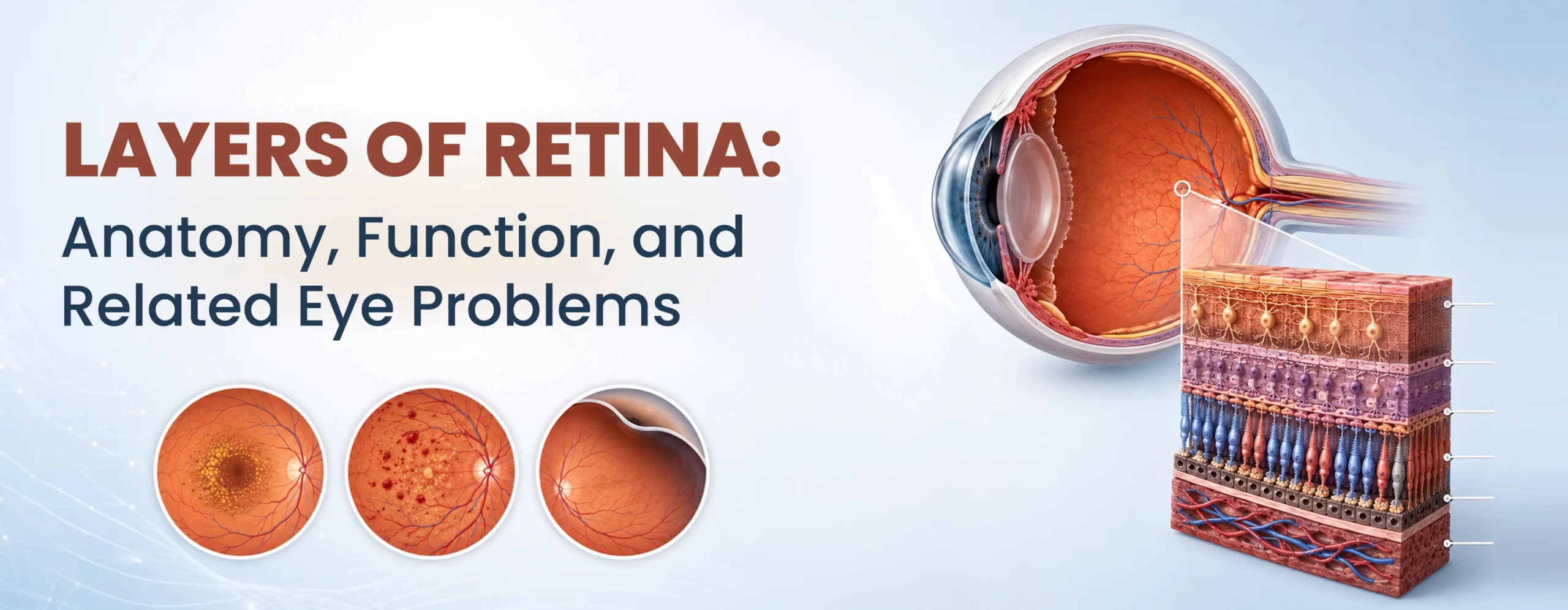
What Are the 10 Layers of the Retina?
Retina specialists describe 10 layers of retina, arranged from the outer surface near your blood supply to the inner surface facing the vitreous gel. Each layer has a distinct job.
A simple way to remember the order, outer to inner:
Really Reds Eat Out Often; Instead, I Generally Need Icecream.
1. R – Retinal Pigment Epithelium (RPE)
A single layer of cells that absorbs stray light and feeds nutrients to your photoreceptors. RPE damage is linked to age-related macular degeneration.
Condition associated – AMD and inherited retinal disorders
2. R – Rods and Cones (Photoreceptor Layer)
This layer of light becomes a nerve signal. Rods manage night and side vision. Cones manage colour and detail.
Condition associated – Retinal degeneration and macular disease
3. E – External Limiting Membrane
A thin structural boundary that holds your photoreceptor cells in place.
Condition associated – Macular and photoreceptor damage
4. O – Outer Nuclear Layer
Contains the cell bodies of your rods and cones.
Condition associated – Retinitis pigmentosa and retinal detachment
5. O – Outer Plexiform Layer
The first point where photoreceptors connect to the next set of nerve cells.
Condition associated – Macular oedema and vascular disease
6. I – Inner Nuclear Layer
Holds cells that refine the signal and help your eyes adjust between bright and dim light.
Condition associated – Retinal vascular and inflammatory disorders
7. I – Inner Plexiform Layer
It passes the signal to ganglion cells.
Condition associated – Vascular and inner-retinal disorders
8. G – Ganglion Cell Layer
Contains your retina’s final output neurons before the signal leaves your eye.
Condition associated – Glaucoma and optic nerve disease
9. N – Nerve Fibre Layer
Gathers ganglion cell fibres and channels them toward your optic nerve.
Condition associated – Glaucoma and optic neuropathy
10. I – Internal Limiting Membrane
The innermost boundary between your retina and the vitreous gel.
Condition associated – Macular hole and epiretinal membrane
Anatomy of the Retina
Where Is the Retina Located?
Your retina lines the inside of your eye, between the vitreous gel at the centre and the choroid, the blood vessel layer behind it. It stretches from just behind your lens to your optic nerve.
What Are the Parts of the Retina?
1. Macula
The small central area responsible for sharp, detailed vision. Its centre, the fovea, gives you your clearest point of focus. Used for reading, recognising faces, and driving.
2. Fovea
The fovea is a small depression at the centre of the macula. It contains a very high concentration of cones and provides the sharpest point of vision.
3. Peripheral retina
The larger area surrounding your macula. Responsible for side vision and low-light vision. Retinal tears frequently develop in the peripheral retina and may be difficult for patients to detect before a detachment begins.
4. Optic Disc
The optic disc is the point where retinal nerve fibres leave the eye and form the optic nerve.
Conditions and Disorders: Common Retina Problems
What Are the Common Eye Diseases That Affect the Retina?
- Diabetic retinopathy – Diabetic retinopathy occurs when prolonged high blood sugar damages retinal blood vessels.
- Age-related macular degeneration (AMD) – Age-related macular degeneration affects the macula, particularly the RPE and photoreceptor complex.
- Retinal detachment and retinal tears – A retinal tear can allow fluid to pass underneath the retina, separating the neurosensory retina from the underlying RPE.
- Retinitis pigmentosa – Retinitis pigmentosa is a group of inherited retinal disorders that progressively damage photoreceptors.
- Macular hole – A macular hole is a small defect in the central retina.
- Epiretinal membrane (macular pucker) – An epiretinal membrane is a thin layer of tissue that forms over the surface of the macula.
- Central serous retinopathy – Central serous chorioretinopathy causes fluid to collect beneath the central retina.
- Retinal vein occlusion – A retinal vein occlusion occurs when a vein carrying blood away from the retina becomes blocked.
A 2022 screening study across 10 Indian states found diabetic retinopathy in 12.5% of adults with diabetes aged 40 and above. Close to 3 million people in India carry a vision-threatening form of the disease.
Also Read: 10 Retina Warning Signs Every Patient Should Know: Advice From Retina Surgeon Dr Aman Kumar
When Should You Consult a Retina Specialist?
Arrange urgent retinal evaluation if you notice:
- A sudden shower of new floaters
- Flashes of light in one eye
- A curtain or shadow moving across vision
- Sudden painless vision loss
- Straight lines appearing bent or wavy
- Loss of peripheral vision
- New visual symptoms after an eye injury
You should also discuss preventive retinal examinations if you have:
- Diabetes
- High myopia
- A family history of retinal detachment
- Previous retinal tears or surgery
- Previous eye trauma
- Known inherited retinal disease
Book an appointment with a retina specialist in Delhi if you notice any of these. Retinal symptoms rarely improve on their own, and some, like a detachment, get harder to treat the longer you wait.
What Are the Retina Treatment Options AKIO Provides?
AKIO is an advanced Retina hospital that offers all services under the roof for retina. Such as –
- Laser photocoagulation for diabetic retinopathy and retinal tears
- Intravitreal injections for AMD, diabetic macular oedema, and retinal vein occlusion
- YAG laser vitreolysis for troublesome floaters
- Vitrectomy for floaters or retinal damage that doesn’t respond to other treatment
- Complete diabetic retinopathy management, from screening to surgery.
Retina Specialists at AKIO
Dr Aman Kumar
Dr Aman Kumar is a Senior Consultant Vitreo-Retinal and Cataract Surgeon at AKIO.
His credentials include:
- AIIMS New Delhi alumnus
- PGIMER Chandigarh Gold Medallist
- Expertise in retinal diagnosis
- Experience in 3D vitreoretinal surgery
- Management of retinal detachment, diabetic retinal disease.
Padma Shri Dr Atul Kumar
Padma Shri Dr Atul Kumar is a highly experienced vitreoretinal surgeon and former Chief of the Dr R.P. Centre for Ophthalmic Sciences at AIIMS, New Delhi.
He brings extensive experience in saving the vision of millions facing retinal disease, macular issues and vitreoretinal surgery.
Together, AKIO’s retina team provides specialist evaluation supported by advanced retinal imaging and surgical technology in Safdarjung Enclave, New Delhi.
Frequently Asked Questions
Can an OCT scan show all 10 layers of the retina?
Modern OCT imaging can distinguish many retinal layers in cross-section, particularly in the macula
Do all eye floaters need treatment?
No. Most harmless floaters require only observation.
How do I take care of my retina?
Get a dilated eye exam once a year, manage blood sugar and blood pressure, and protect your eyes from UV light.
Can damaged retinal layers be repaired?
Some damage can be treated with lasers, injections, or surgery, but retinal nerve cells don’t regenerate once lost.
What is the first warning sign of retinal disease?
Sudden floaters, flashes of light, blurred vision, distortion, or a curtain-like shadow may all be early signs of retinal disease.
Can stress damage the retina?
Stress itself does not directly damage the retina, but it has been associated with conditions such as Central Serous Chorioretinopathy (CSCR) in some patients.
Can retinal disease occur without symptoms?
Yes. Diabetic retinopathy and some peripheral retinal changes may develop before patients notice vision problems.
How often should people with diabetes get a retina examination?
People with diabetes should undergo a dilated retinal examination at the interval recommended by their ophthalmologist. Annual assessment is commonly advised, but more frequent follow-up may be necessary when diabetic retinopathy is present.
How often should we examine our eyes for good retinal health?
Once a year for most adults, and every 6 months with diabetes or high blood pressure.


Recent Posts
- How to Choose the Right IOL for Cataract Surgery: A Patient’s Guide
- Can A Retinal Tear Heal On Its Own? When Is Retinal Laser Needed?
- Macular Hole: Why Straight Lines Look Bent, or Faces Look Distorted
- Can Retinal Detachment Cause Permanent Blindness? How Fast Should You Get Treatment?
- Eye Floaters – When Are They Normal And When Could They Be A Sign of Retinal Detachment?